
Tags
Related Posts
Share This


Nanocodes: Imaging on Molecules
by Henry Freedman
Nano marking, already in use in scientific lab research, carries implications for commercial printing. It is strikingly similar to ion deposition printing technology developed by Delphax.
Just as production environments in photo finishing, printing and publishing have workflow tracking requirements, so do areas of scientific research in biochemistry and medicine. Commercial imaging and security printing industries may one day benefit from emerging medical nano marking already in use in laboratory settings. Medical researchers can now mark, and track molecules by recording what are called nanobarcodes” on the molecules themselves. These molecules are sub-micrometer in size. This shows how far we have come placing marks on substrates.
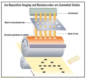
Using a sequential electrodeposition process, metal ions, typically of gold and silver, are deposited on molecules, called “microrods,” by transmitting the ions through a template that has an array of holes or pores. This appears not to be too far from a device familiar in digital printing circles, the Delphax ion deposition printing technology. This system, commercialized in 1981, used an approach that tunneled electrons through a mask to create a printed dot. For nanobarcodes, the contrast between the gold and silver stripes on the molecules allows optical reading of the printed line on the molecule.
New Language for a Line
Since the scientific community is not necessarily versed in the language used for printed images, we find new terminology popping up. For example, research papers use the term “submicrometer stripe” to refer to what we know as a recorded barcode line. What is also most interesting is the great similarity of the nanobarcode process to letterpress electroplating and to chemical etching of relief plates. Also of note is that the process may actually be another form of sublimation imaging, that is, imaging within the substrate.
Reading the Nanobarcode
In the lab applications, the striping pattern is read out via optical microscopy, typically by imaging the reflectivity of the particles under blue light illumination. Silver is highly reflective under blue light and gold is not, leading to a bright/dark pattern on the alternating silver (Ag) and gold (Au) striped particles. So as you can see, the barcode pattern is an intrinsic feature of the particles themselves. The particles can be used to encode the identity of a molecule (e.g. a protein or DNA sequence), adsorbed to the surface of the particles. Note that these particles are on the order of 6 microns in length and 300 nm in diameter. While they are very small, they are much larger than the molecules they are used to identify.
Thousands of biomolecules can be attached to the outside of each single particle. This is sometimes hard for people to envision, as one would expect the barcode to be a small tag placed onto a larger object (like we see with conventional barcodes in the retail industry).